You may or may not remember, but a while back a video entitled "The Inner life of the cell" was posted on PPT. The video was made by Biovisions at Harvard University and can be found
Since a number of people commented at the time that they would like to know what was going on in the video, I thought I would break it down and explain it!
|
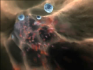
We start in the middle of a blood vessel. The red things you see flying by are the red blood cells (Erythrocytes), and the blue-coloured cells moving more slowly along the edge of the vessel are white blood cells.
|

|
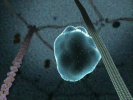
Next we zoom in to see the interactions between the vessel wall and the white blood cell - this one is called a Leukocyte. It is "crawling" along the vessel wall - later on in the video we will discover why.
|
|
Here we have zoomed in once again and can see how the Leukocyte is holding onto the vessel wall. Proteins on the cell and the vessel wall attach to each other to adhere (or stick) together. The strength of these interactions isn’t very strong but when there are a whole load of them, as there are on the cell, it’s very strong - much like all the little hooks on Velcro - they’re weak on their own, but when there’s lots it’s really strong.
|
|
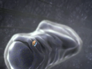
Moving on in the video we zoom in once again, to see the outer side of the lipid bilayer that forms the membrane of the cell. On the lipid bilayer there are many proteins which transmit messages from the outside of the cell to the inside.
|
|
We have now moved from outside of the cell to the inside where we can see there are proteins attached to the inside of the membrane. You can also see the cytoskeleton, which is what holds cells in the shape they need to be in (cyto meaning cell, and the skeleton has the same function as the skeleton made of bones that supports your body).
|
|
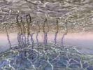
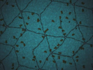
We have now moved away from the membrane and looked back up towards the membrane. Groups of a protein called Spectrin are holding the filaments of another protein called actin together like scaffolding maintaining the shape and strength of the membrane.
|

|
Now we can see the rest of the cytoskeleton further into the cell. It is made of filaments of Actin held together by Actin Binding poteins.
|
|
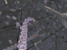
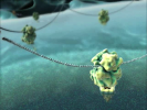
Here we can see how an Actin filament forms - it is made out of actin monomers, which are little globular (or blob shaped) proteins, which spontaneously polymerise and depolymerise (join and leave the filaments) at the leading end depending on the amount of monomers in the area. Actin is a very dynamic structure, constantly being remodelled.
|

|
When the cell wants to break the Actin filament a severing protein can break the filament in a specific place by forcing the filament into a kink which breaks the bonds, breaking the Actin filament at the desired spot.
|
|
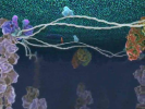
Another component of the cytoskeleton are Microtubules. They are made of Protofilaments which are in turn made from Tubulin dimers. Like Actin, Microtubules are dynamic structures which are being made and broken down all the time.
|
|
|
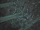
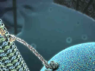
Mictotubules are a bit like the roads in a cell - they are paths that motor proteins, in this case probably Kinesin, can walk along.
|
|
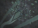
The inside end of the Microtubules are anchored by the centrosome, which stabilises them giving them more of a structure.
|
|
|
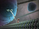
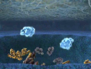
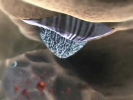
Now we have moved down just outside of the nucleus. The nuclear membrane it’s a specialised membrane and contains structures called nuclear pores, which you can see in this picture. mRNA and its associated proteins exit the nucleus through these nuclear pores.
|
|
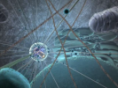
In the cytoplasm the mRNA associates with ribosomes, which translate the message contained on the mRNA into a protein.
|
|
|
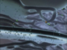
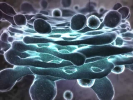
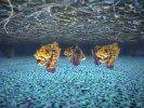
Here we can see a Mitochondria. Mitochondria are the "power houses" of a cell. They are vitally important in the conversion of energy from the food we eat into a form the cell can use, which is predominantly ATP. Mitochondria are a special type of organelle, as they contain DNA, the only place you find DNA outside the nucleus.
|
|
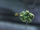
Proteins that will be secreted from the cell or the proteins that are integrated into the cell membrane are made in the Endoplasmic Reticulum (often shortened to ER). Vesicles, which are little bubbles of membrane, then bleb (yes that is a real word!) off the ER and are moved up the Microtubule tracks by the motor proteins.
|
|
|
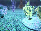
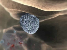
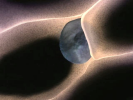
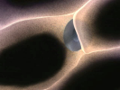
The next stop is the Golgi Apparatus. The vesicle fuses with the golgi and the proteins contained within it are processed, for example glycosylated, which means a sugar group is added to them. Some proteins require this processing before they are fully functional.
|
|
Once the proteins are fully processed and ready to go the vesicle is transported up the Microtubule to the cell membrane, as we saw at the beginning of the video.
|
|
When it reaches the membrane the vesicle fuses with the membrane and the proteins to be secreted are let out into the extracellular space (the space outside the cell). The membrane bound proteins can then diffuse around the cell membrane to where they need to be. Here you can see the lipid rafts form that we saw earlier. These rafts are special areas of the membrane that are made of a slightly different kind of lipid and have more cholesterol between the lipid molecules. The lipid rafts in the video contain Integrins.
|
|
|
The Integrins on the cell surface respond to the presence of Cytokines on the vessel wall. Cytokines are a molecule which is created in inflamed tissue. The detection of these molecules tells the Lymphocyte that there is inflammation in the tissue behind the vessel wall. This triggers a pathway inside the cell that causes the cytoskeleton to change dramatically and the cell squeezes between the endothelial cells (the cells that make the vessel wall) to enter the tissue and help the body fight off the infection causing the inflammation.
|
|
has a rich inner life.


